Waterjet endoscope 有幫助清除 blood clots
Suction 不可
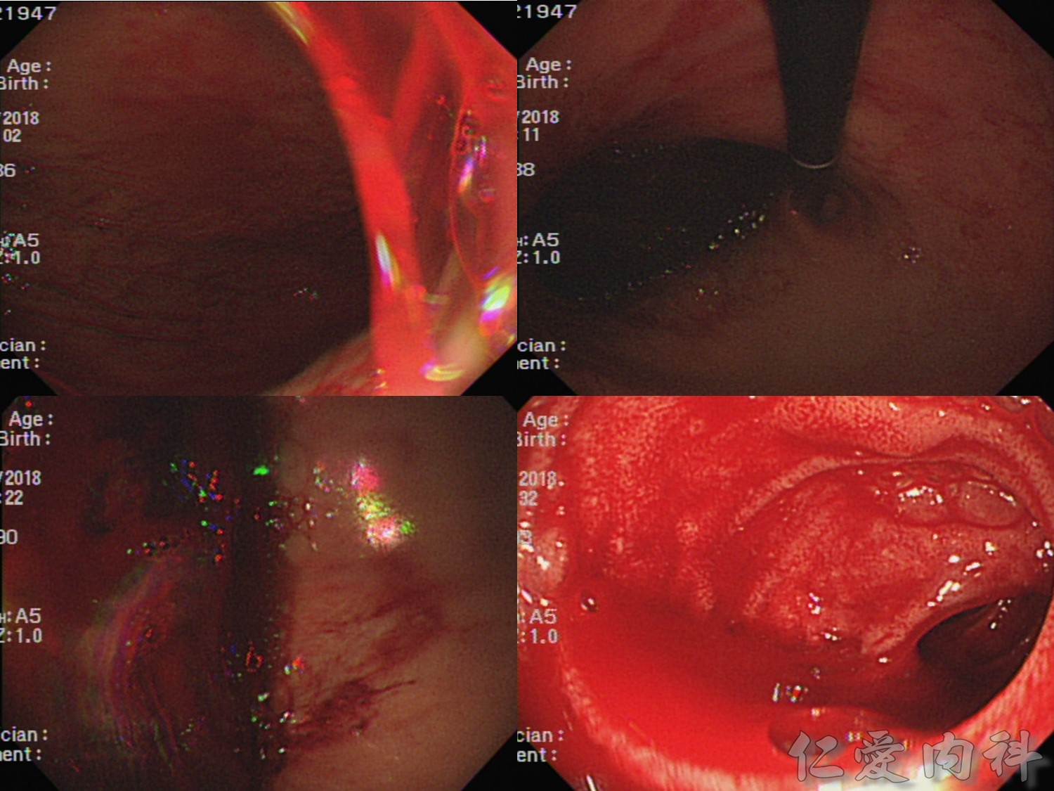
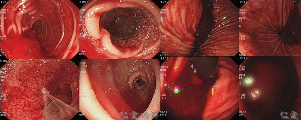
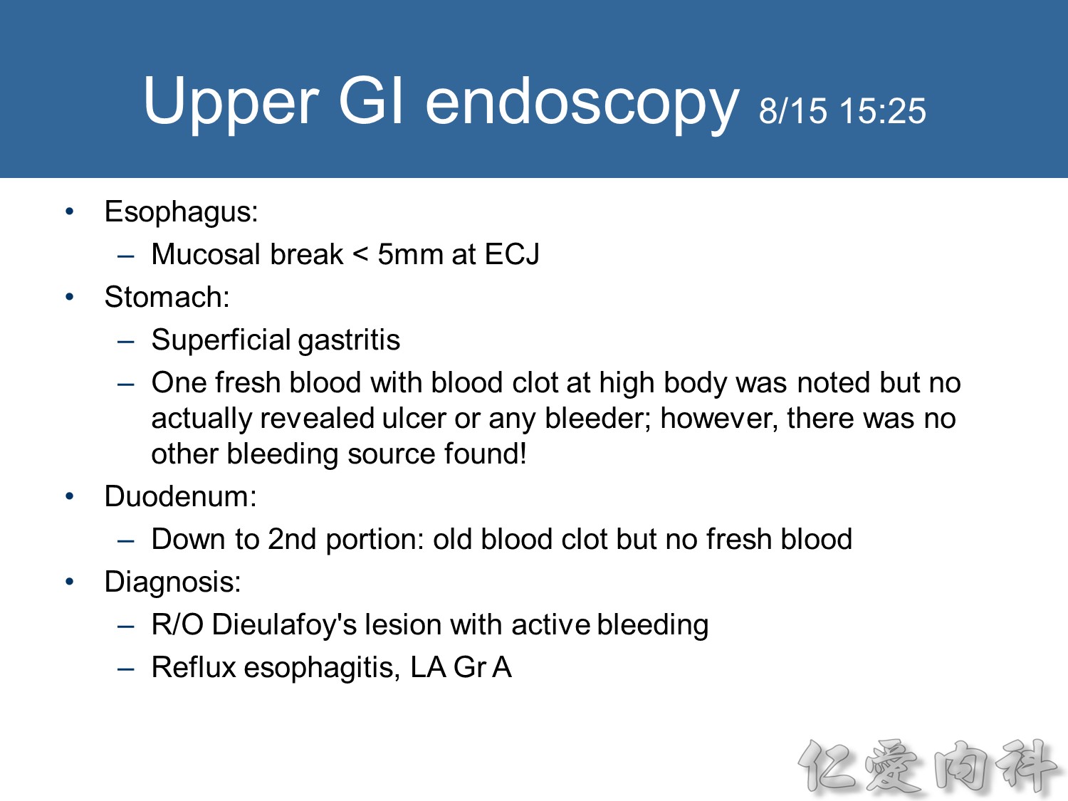
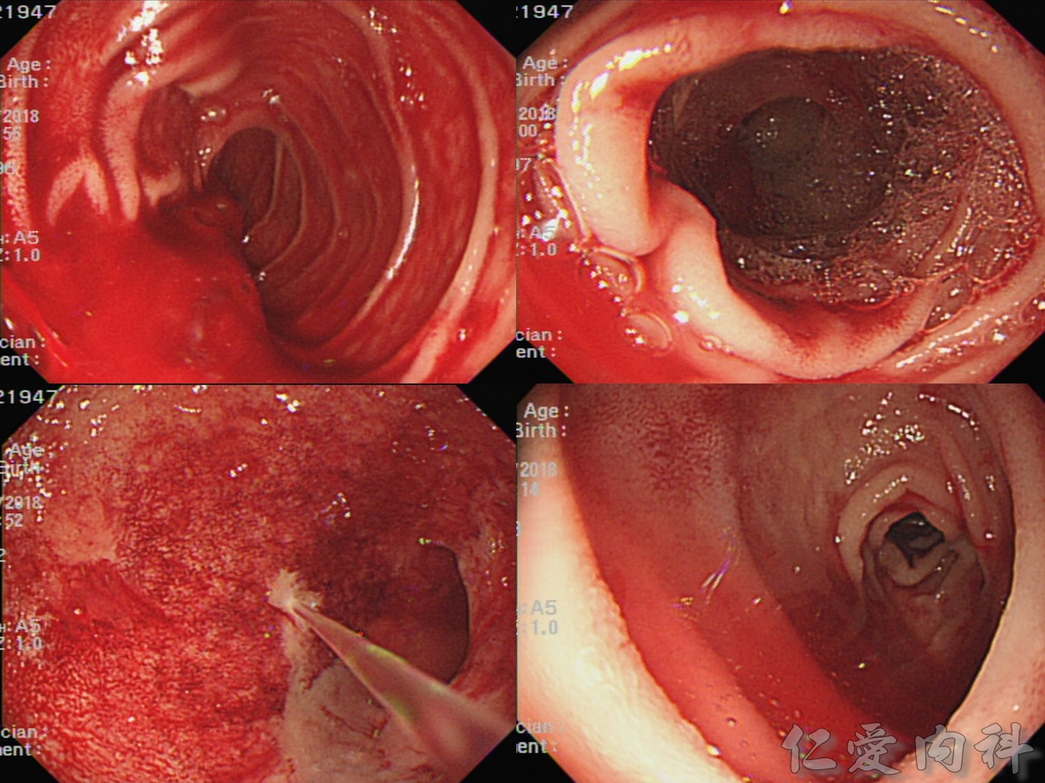
第二次胃鏡進去在 cardia 部分一開始是看到黑、紅交雜的新舊 blood,進去在 duodenum 的地方還是可以看到 fresh blood。我們沖了很多次水後基本上 duodenum 就清的蠻乾淨,看不到 fresh blood 了。但是勾回來看還是看的到 cardia、fundus 的地方有黑紅色的血冒出,高度懷疑是 high body bleeding。
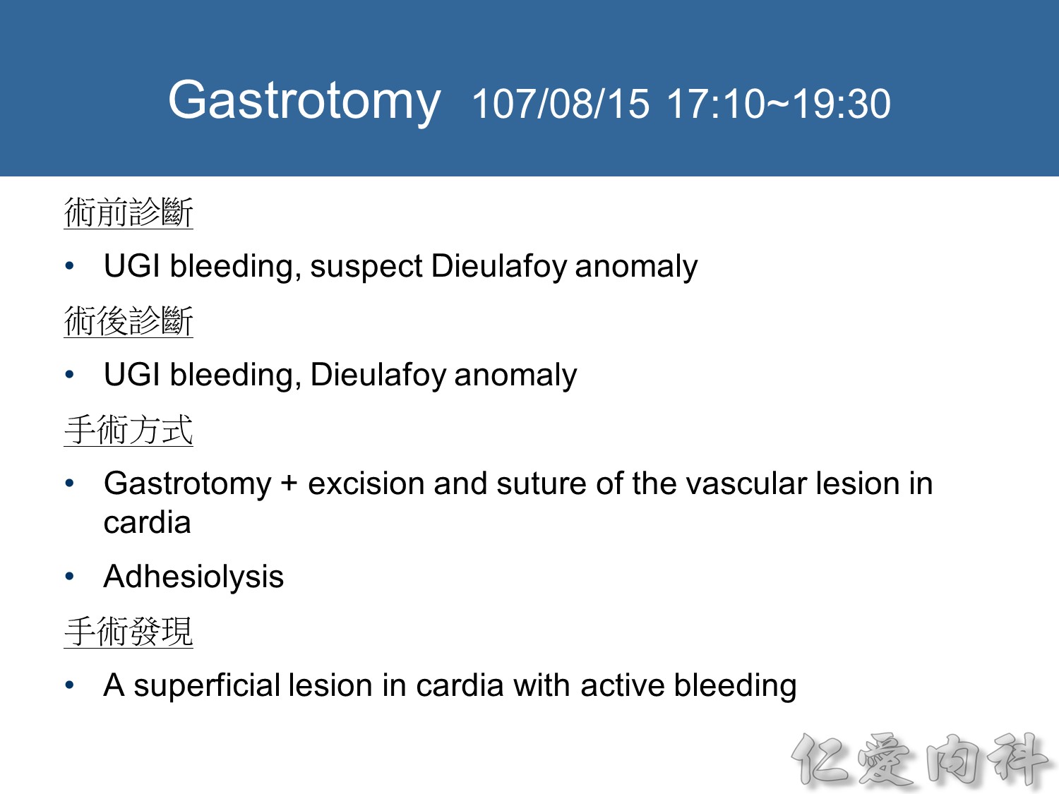
手術過程
1. Position: supine
2. Incision: upper midline
3. Divided the subcutaneous tissue and the fasciae layer.
4. Opened the peritoneum and entered the abdominal cavity. There were diffuse adhesions betweent the peritoneal wall and the stomach, colon and intestines.
The adhesions were divided with electrocauterization and sharp dissection.
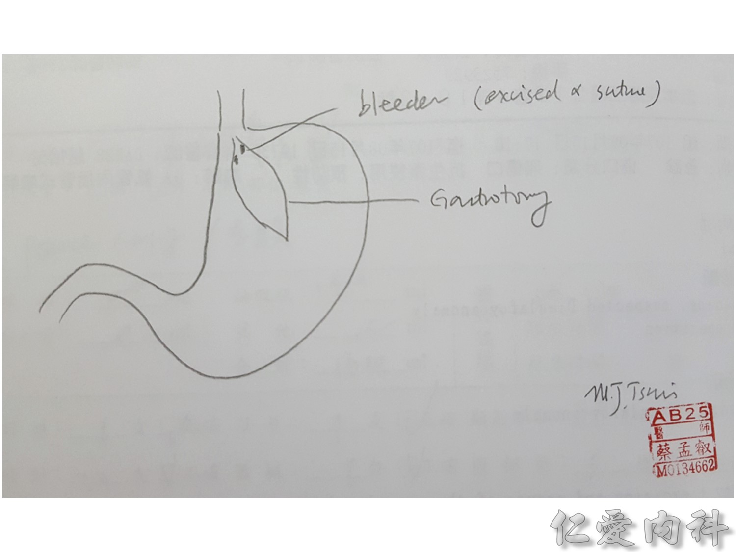
5. A large incision was made on the anterior wall of the stomach to near the cardia.
6. A shallow lesion was found in the cadia near the esophageocardia junction.
The lesion was excised and the defect was closed primarily.
7. The high body of the stomach and fundus were checked again. No other lesions with bleeding were found.
8. Another incision was made in the antrum to near the pyloric area, and a shallow ulcer was found in the duodenum, first portion. The ulcer was suture with silk 000.
9.The lower gastrotomy wound was closed.
10.The upper stomach was checked again. No vascular lesion with bleeding was found.
11.The gastrotomy wound was closed with Endo-GIA.
12.Two penrose drains were placed in the left subphrenic space.
13.The laparotomy wound was closed in layers.
14.The patient was sent to the surgical ICU for monitoring and intensive care.